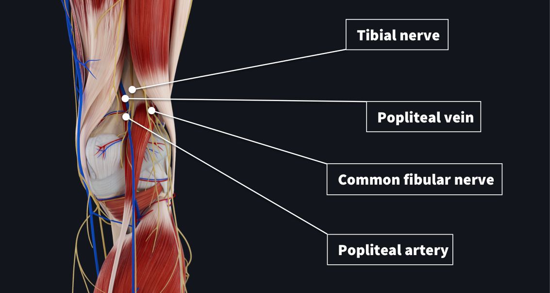
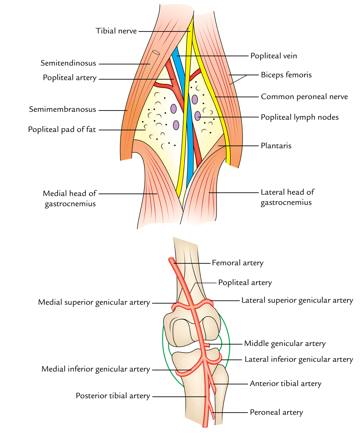
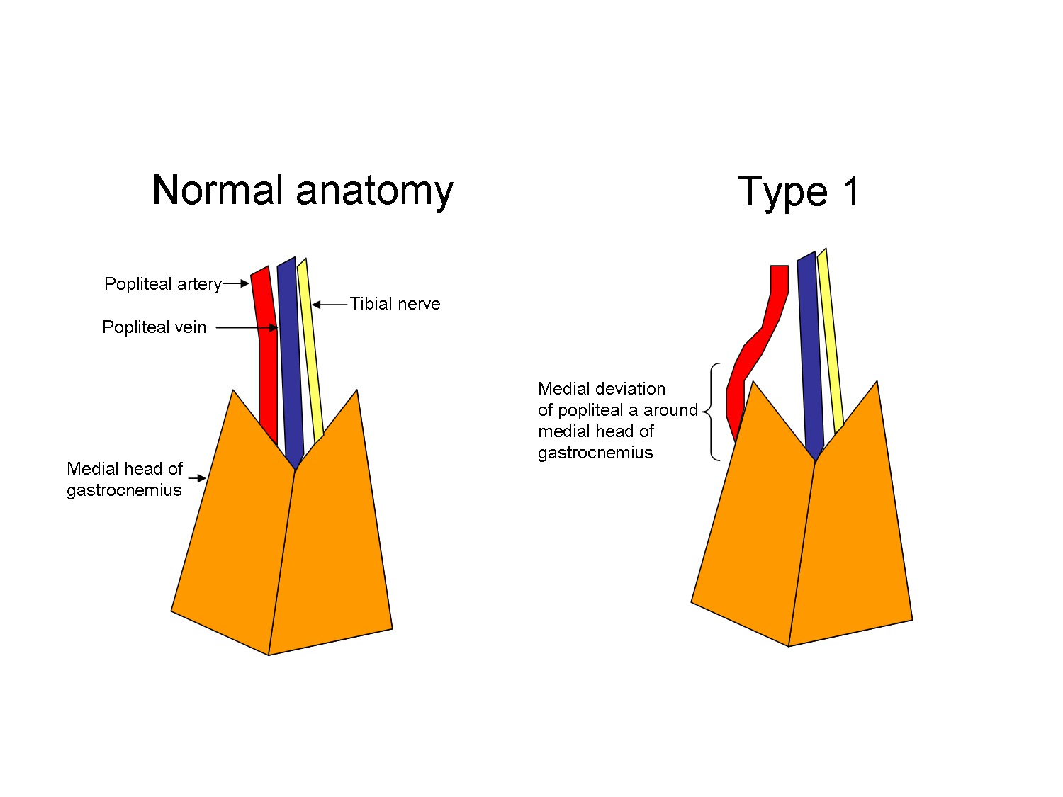
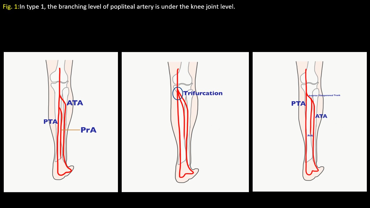
In close proximity to the artery, within the confines of the popliteal fossa, are the tibial nerve, common peroneal nerve, and the popliteal vein An understanding of the normal anatomy and the important variations in the popliteal bifurcation patterns is essential In this report, we have combined data from new cadaver dissections with prior anatomical data to describe theMost anatomy textbooks briefly describe a single popliteal vein, and the literature contains few references on venous patterns in this region Although the primary objective of this study was to analyze venous variability in 52 dissected cadaveric popliteal fossae and 63 venograms, data were also collected on the popliteal artery Nine groups (AI) were designated regarding the mannerYou can use the
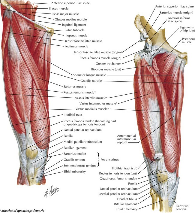
Exposure Of The Popliteal Artery And Vein Basicmedical Key
Popliteal artery vein anatomy
Popliteal artery vein anatomy- Chapter 38 Exposure of the Popliteal Artery and Vein Matthew T Allemang and Vikram S Kashyap Introduction Exposure of the popliteal artery and vein is relevant to many surgical specialties, including general, vascular, and trauma The approach to the popliteal anatomy can be dictated by the pathology, with specific conditions unique to this mobile areaA STUDY OF POPLITEAL ARTERY AND ITS VARIATIONS WITH CLINICAL APPLICATIONS Submitted in partial fulfillment for MD DEGREE EXAMINATION BRANCH XXIII, ANATOMY Upgraded Institute of Anatomy Madras Medical College and Rajiv Gandhi Government General Hospital, Chennai 600 003 THE TAMILNADU DrMGR MEDICAL UNIVERSITY CHENNAI – 600


Nerve Knee Muscle Popliteal Fossa Popliteal Artery Popliteal Artery Angle Text Human Png Pngwing
Exposure of the popliteal artery and vein is relevant to many surgical specialties, including general, vascular, and trauma The approach to the popliteal anatomy can be dictated by the pathology, with specific conditions unique to this mobile area Thus, this chapter encompasses both medial and posterior approaches to the popliteal artery and Key words popliteal vein, sciatic vein, anatomy variations INTRODUCTION Venous anatomy is known to be highly variable In the lower limbs the most commonly present are two systems collecting blood deep and superficial They are separated by the fascia surrounding the thigh muscles, which defines the two compartments of the thigh the superficial compartment, femoral artery vein nerve anatomy 2591Femoral artery vein nerve anatomy リンクを取得 ;
The popliteal vein is formed by the junction of the venae comitantes of the anterior and posterior tibial vein at the lower border of the popliteus muscle It travels on the medial side of the popliteal artery As it ascends through the fossa, it crosses behind the popliteal artery so that it comes to lie on its lateral sideMale reproductive system Testicles;One lies beneath the popliteal fascia near the termination of the external saphenous vein, another between the popliteal artery and the back of the kneejoint, while the others are placed at the sides of the popliteal vessel Arising from the artery, and passing off from it at right angles, are its genicular branches
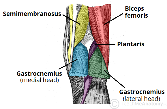
The popliteal fossa is a diamond shaped area located on the posterior aspect of the knee It is the main path by which vessels and nerves pass between the thigh and the leg In this article, we shall look at the anatomy of the popliteal fossa – Dr Hadzic demonstrating the relevant anatomy for the popliteal block on an ultrasound image In the sheath of the sciatic nerve, we can find the tibial nerve and the common peroneal nerve These two nerves are always enclosed in Vloka's sheath, and underneath you have the popliteal vein which is more superficial, and the popliteal artery The one thing that youInferior thyroid vein Article Media The inferior thyroid vein (Latin vena thyroidea inferior) is a blood vessel that arises from


Basic Anatomy Of The Lower Extremity Arteries Medmastery


Knee Page 2 Anatomy Exhibits
Female reproductive system Ovaries; Popliteal fossa artery vein nerveIn anatomy, a hollow or depressed area amygdaloid fossa the depression in which the tonsil is lodged cerebral fossa any of the depressions on the floor of the cranial cavity condylar fossa ( condyloid fossa ) either of two pits on the lateral portion of the occipital The tibial nerve is first lateral to theAnatomy of female body with arteries and veins popliteal artery stockgrafiken, clipart, cartoons und symbole old engraved illustration of first aid when cutting the popliteal artery popliteal artery stockfotos und bilder


Popliteal Artery Entrapment Syndrome Paes


Humannervesspinalfoot Ahuman Human Spinal Lumbosacral Nerves Foot And Lower Leg Create The Personality Nerve Anatomy Anatomy Sciatic Nerve
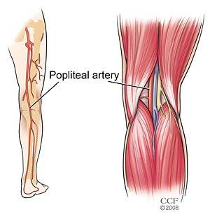
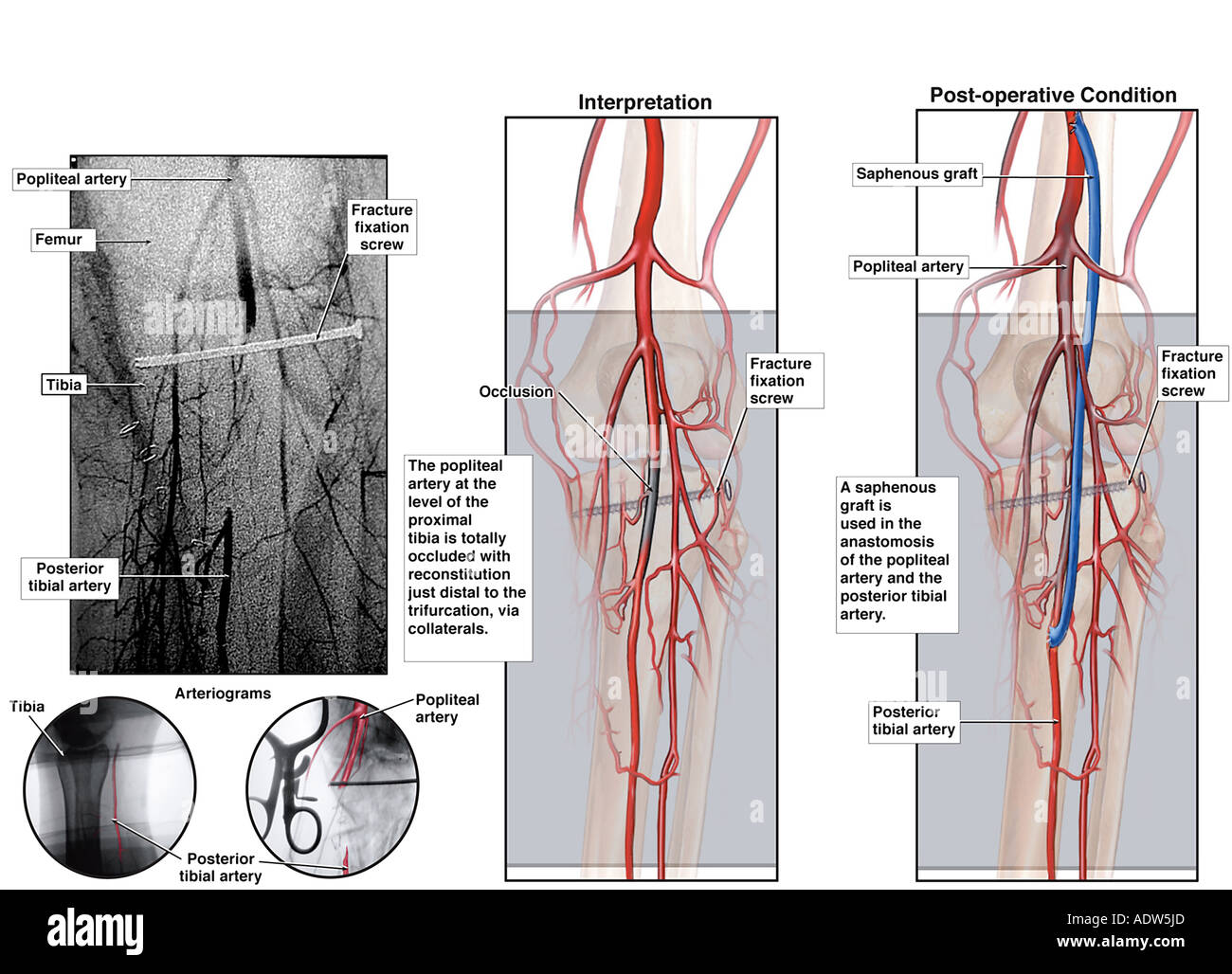
Popliteal artery entrapment release involves open surgery to reestablish normal musculature anatomy behind the knee, and repair chronic damage to the popliteal artery Mr Robert Davies is a recognised UK expert in popliteal entrapment syndrome attracting work from around the country The popliteal vein is located posterior to the knee in the popliteal region that is a major route for venous return from the lower leg The vein forms from the combination of the anterior and posterior tibial vein at the border of the popliteal artery The vein is found in the popliteal fossa on the posterior aspect of the knee The vein crosses from the medial to the The aboveknee popliteal artery starts at the distal adductor canal (where the thigh becomes the knee), and the belowknee popliteal artery extends to the bifurcations of the calf arteries at the distal popliteal fossa The popliteal is the only artery where you regularly see the vein located above the artery on the ultrasound screen Figure 6


Popliteal Artery High Resolution Stock Photography And Images Alamy


Popliteal Artery
Popliteal vein the small saphenous vein passes between the two heads of gastrocnemius to empty into popliteal vein ; Popliteal artery– deepest structure, continuation of femoral artery ;Tibial nerve– most superficial, branch of sciatic nerve ;


Figure Popliteal Lymph Nodes Tibial Nerve Statpearls Ncbi Bookshelf


Nerve Knee Muscle Popliteal Fossa Popliteal Artery Popliteal Artery Angle Text Human Png Pngwing
9月 19, 21 The femoral artery is the continuation of the external iliac artery in the thigh becoming the femoral artery as it passes under the inguinal ligament TheA little above the knee the popliteal vessels are joined by the sciaticThe popliteal lymph glands, six or seven in number, are imbedded in the fat;The anatomic relationship between the popliteal artery and vein means that an arteriovenous fistula can be created when a popliteal artery approach is used for endovascular interventions To determine the best site for retrograde puncture of the popliteal artery, we studied six cadaveric specimens, CT scans of 31 patients at 280 levels, and 30 plain radiographs of the knee In the


The Popliteal Fossa Borders Contents Teachmeanatomy


Progressive Stenosis Of A Popliteal Artery Stent Graft By Laminated Thrombus Journal Of Vascular Surgery Cases Innovations And Techniques
The anatomic relationship between the popliteal artery and vein means that an arteriovenous fistula can be created when a popliteal artery approach is used for endovascular interventions To determine the best site for retrograde puncture of the popliteal artery, we studied six cadaveric specimens, CT scans of 31 patients at 280 levels, and 30 plain radiographs of the knee In theOld engraved illustration of first aid when cutting the popliteal artery popliteal artery stock pictures, royaltyfree photos & images female anatomy of cardiovascular system, rear and front views popliteal artery stock illustrations Illustration from 'Surgical Anatomy The Treatise of the Human Anatomy and Its Applications to theMRI of the knee is often performed for presumed musculoskeletal conditions There is a wide variety of variant vascular anatomy and vascular pathology that can occur around the knee, including an aberrant anterior tibial artery, vascular trauma that occurs with knee dislocation, popliteal artery entrapment syndrome, popliteal artery aneurysm, popliteal vein thrombosis, cystic adventitial


Arteries And Veins Of The Lower Limb Dr


Exposure Of The Popliteal Artery And Vein Basicmedical Key
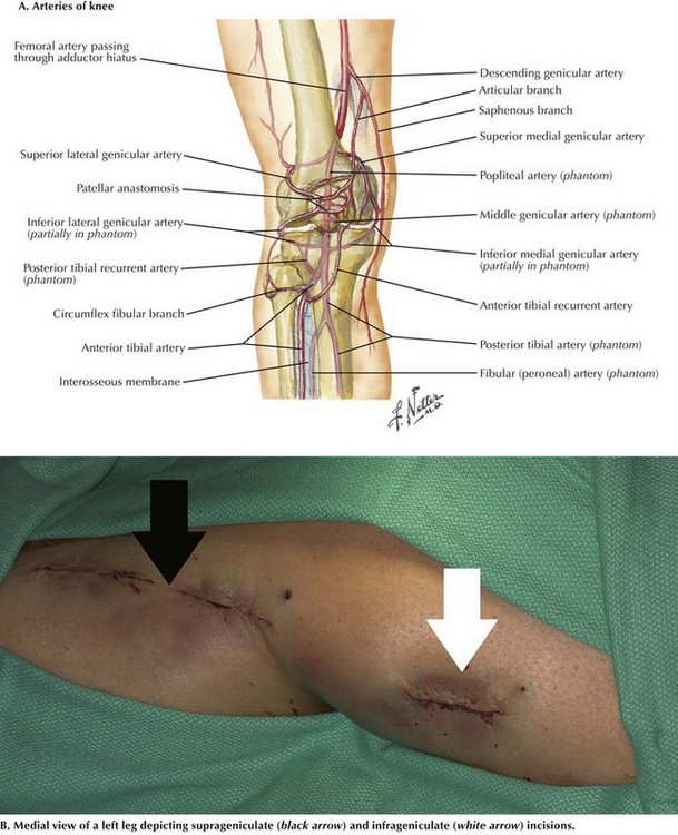
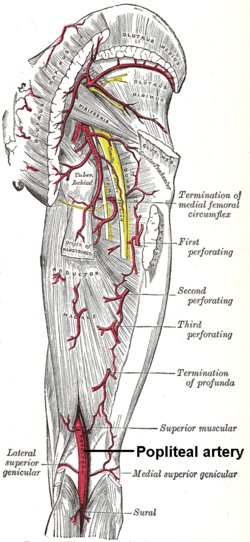
Popliteal artery is the extension of femoral arteryThe starting point is adductor hiatus (an opening of osseoaponeurotic type located in adductor magnusat , the junction of middle onethird and lower onethird of thigh), it gets divided into anterior and posterior tibial arteries when it comes across the floor of popliteal fossa by the medial to lateral side to reach the border of the popliteusElementary anatomy and physiology for colleges, academies, and other schools A View of the Arteries on the Back of the Leg The Muscles have been removed so as to display the Vessels in their whole length 1, The Popliteal Artery, cut off so as to show the Articular Arteries 2» 375Dr Ebraheim's educational animated video describes the anatomy of the back of the knee Popliteal Fossa The area of depression located at the back of the


Popliteal Artery Entrapment A Mysterious Syndrome


Thigh Knee And Popliteal Fossa Knowledge Amboss
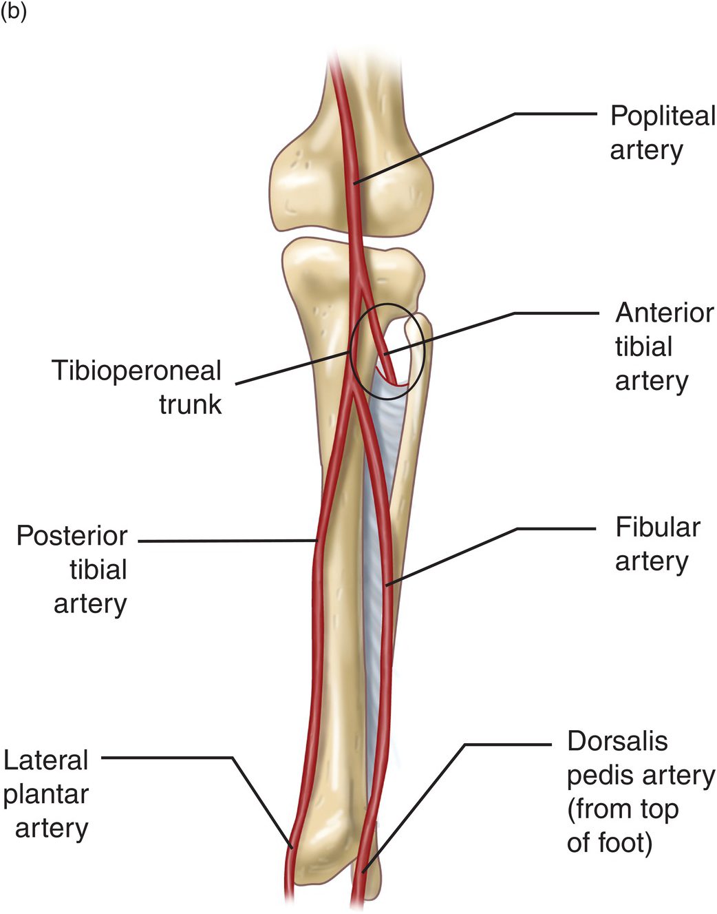
The Popliteal Artery branches & genicular anastomosis The Popliteal Artery branches & genicular anastomosis Watch later Share CopyThe popliteal artery is a continuation of the superficial femoral artery as it passes through the adductor hiatus of the adductor magnus muscle, traveling posteriorly to the knee and anterior to its accompanying vein, the popliteal vein, until it bifurcates into the anterior tibial artery and the common trunk of the posterior tibial and peroneal arteries Geniculate arteries branch atThe popliteal vein arises at the lower border of the popliteus muscle, ascends through the popliteal fossa and passes through the adductor canal, becoming the femoral vein The main tributaries of the popliteal vein are the small saphenous vein, the muscular veins and the veins corresponding to the branches of the popliteal artery The popliteal vein collects blood from the knee joint, as


Leg Popliteal Artery Damage


Popliteal Fossa Bf Biceps Femoris Pa Popliteal Artery Pv Download Scientific Diagram
Joins the posterior tibial vein The popliteal vein is formed by the junction of the anterior and posterior tibial veins at the lower aspect of the posterior knee It ascends along the posterior aspect of the knee and the distal aspect of the anteromedial thigh The popliteal vein is located medial to(330 Ligation of the popliteal artery below the inner condyle of the tibia (S6dillotJ nerve, the popliteal vein, and the artery The nerve and vein are to be drawninward, and the needle passed from within outward (3) To ligature the popliteal artery below the internal condyle of the tibia,semillex the leg, and lay it upon the outer side Popliteal artery entrapment syndrome (PAES) occurs when muscles that surround the popliteal artery in the area of the popliteal fossa, occlude the artery (and sometimes the vein as well), and decrease blood flow to the lower leg Two forms of PAES exist anatomical and functional Figure 1 Typical anatomy of the popliteal fossa


Safe Zones For Pin Placement


The Popliteal Fossa Complete Anatomy
Lymphatic system Spleen ;Veins of pelvis and lower limb;The popliteal vein may be deeper than the artery or separated from it by a slip of the gastrocnemius muscle (a third head of the gastrocnemius) A third head of the gastrocnemius is the most common variation of the gastrocnemius muscle and there are several varieties of the third head These variations of the gastrocnemius may compromise the function of one or more of the


Popliteal Fossa Fossa Common Fibular Nerve Anatomy


Popliteal Vein Wikipedia
The popliteal artery is a continuation of the superficial femoral artery as it passes through the adductor hiatus of the adductor magnus muscle, traveling posteriorly to the knee and anterior to its accompanying vein, the popliteal vein, until it bifurcates into the anterior tibial artery and the common trunk of the posterior tibial and peroneal arteries Geniculate arteries branchCommon fibular nerve– most superficial, branch of sciatic nerve and travels along lateral margin of fossa ; Arteries (right and left hepatic artery branches) Vein (portal vein) Epiploic foramen (foramen of Winslow) Femoral triangle or Scarpa's triangle Mnemonic NAVEL From lateral to medial Nerve (femoral nerve and femoral branch of genitofemoral nerve) Artery (femoral artery) Vein (femoral vein and it's tributary – great saphenous vein)


Easy Notes On Popliteal Fossa Learn In Just 4 Minutes Earth S Lab


Http Medinfo2 Psu Ac Th Surgery Collective review 2559 26 Popliteal Artery Aneurysms Paas Chakwit 21 12 59 Pdf
Surgical Anatomy of the Popliteal Vessels The popliteal artery is a short but vital segment of the major arterial conduit of the leg situated between the adductor hiatus and the lower border of the popliteus muscle posterior to the knee joint ( Fig 171 ) Fig 171 The popliteal artery extends from the adductor hiatus to the lower border ofThe popliteal artery (Fig 551) is the continuation of the femoral, and courses through the popliteal fossa It extends from the opening in the Adductor magnus, at the junction of the middle and lower thirds of the thigh, downward and lateralward to the intercondyloid fossa of the femur, and then vertically downward to the lower border of the Popliteus, where it divides into anterior andMuscles of Leg (Intermediate Dissection) Posterior View Anatomy Adductor magnus tendon, Popliteal artery (deeper) and vein (more superficial), Superior medial genicular artery, Tibial collateral ligament, Semimembranosus tendon (cut), Inferior medial genicular artery, Popliteus muscle, Plantaris tendon, Gastrocnemius muscle (cut), Flexor digitorum longus tendon, Tibialis


Thumb Knee Femoral Artery Popliteal Artery Crus Others Angle Hand Foot Png Pngwing


Popliteal Artery Wikipedia
The popliteal artery located behind the knee is where the popliteal vein begins to extend Popliteal vein begins at the lower border of the popliteus by the union of veins accompanying the anterior and posterior tibial arteries Popliteal Artery During its course the popliteal artery branches into other significant blood vessels Popliteal anatomy The bones of the popliteal
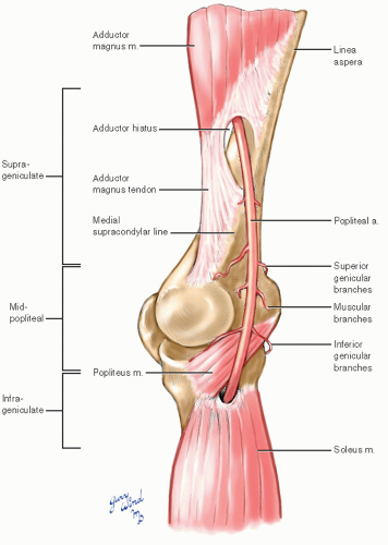


Popliteal Artery Basicmedical Key


Popliteal Artery Entrapment Syndrome In A Young Baseball Pitcher A Ca Jpr


Popliteal Artery Dr Ahmed Farid Youtube
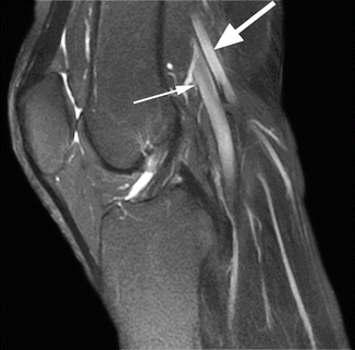


Arteries And Nerves Radiology Key


Vascular Problems Of The Knee Musculoskeletal Key


Safe Zones For Pin Placement


Popliteal Artery Location Entrapment Popliteal Artery Aneurysm


Popliteal Sciatic Nerve Block Landmarks And Nerve Stimulator Technique Nysora


A Current Interpretation Of Popliteal Vascular Entrapment Sciencedirect


Http Medinfo2 Psu Ac Th Surgery Collective review 2559 26 Popliteal Artery Aneurysms Paas Chakwit 21 12 59 Pdf


3 10 Popliteal Fossa And Leg Flashcards Quizlet


A Rare Cause Of Popliteal Artery Entrapment Syndrome Ejves Extra


Jaypeedigital Ebook Reader


Thigh Knee And Popliteal Fossa Knowledge Amboss
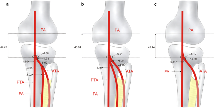


Variations In Terminal Branches Of The Popliteal Artery Cadaveric Study Springerlink


Exposure Of The Popliteal Artery And Vein Basicmedical Key


1


Popliteal Artery Entrapment A Mysterious Syndrome
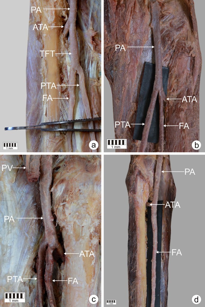


Variations In Terminal Branches Of The Popliteal Artery Cadaveric Study Springerlink


Surface Marking Of Popliteal Vein Download Scientific Diagram


7 Figure Settlement Surgeons Damage Blood Vessels During Total Knee Replacement High Impact Visual Litigation Strategies


Figure Posterior View Of The Nerve Statpearls Ncbi Bookshelf


Epos C 1736


Right Popliteal Vein Aneurysm A Case Report


Epos


Popliteal Anatomy Definition Anatomy Drawing Diagram


Figure 1 From Anatomic Variations Of Popliteal Artery That May Be A Reason For Entrapment Semantic Scholar


Popliteal Fossa Radiology Reference Article Radiopaedia Org


1


6 Anatomy Of Popliteal Fossa


Popliteal Artery Chapter 36 Atlas Of Surgical Techniques In Trauma


1
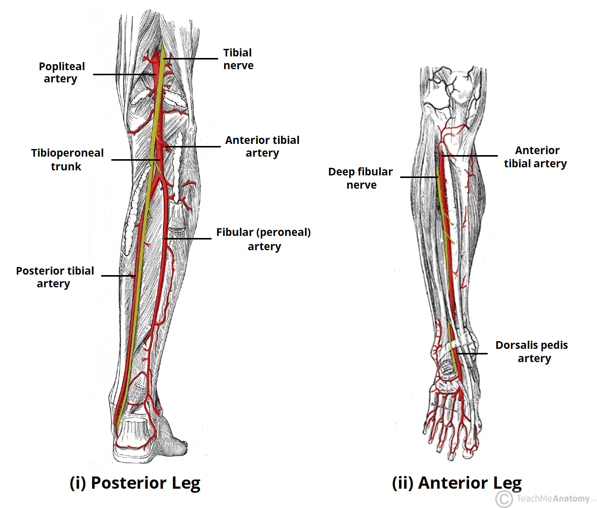


Arteries Of The Lower Limb Thigh Leg Foot Teachmeanatomy
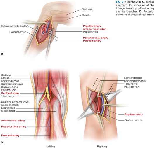


Surgical Exposure Of The Lower Extremity Arteries Thoracic Key
:watermark(/images/watermark_only.png,0,0,0):watermark(/images/logo_url.png,-10,-10,0):format(jpeg)/images/anatomy_term/vena-poplitea/1qimxsaGtAeyhFKKAmU4Qw_bzAdvntzTh_Vena_poplitea_1.png)


Popliteal Fossa Anatomy And Contents Kenhub
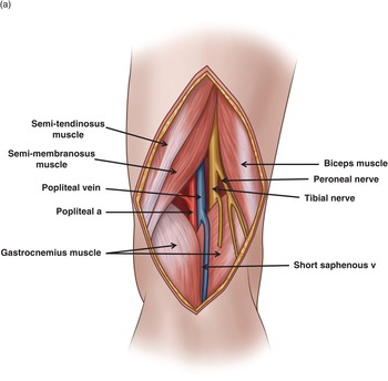


Popliteal Artery Chapter 36 Atlas Of Surgical Techniques In Trauma


Popliteal Artery
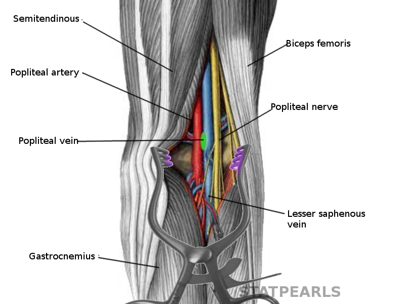


Anatomy Bony Pelvis And Lower Limb Popliteal Vein Article


Thigh Popliteal Fossa Yeditepe Anatomy Lab
:watermark(/images/watermark_only.png,0,0,0):watermark(/images/logo_url.png,-10,-10,0):format(jpeg)/images/anatomy_term/popliteal-artery-2/2s1uAQS90eE45BGW3ovaw_A._poplitea_02.png)


Popliteal Artery Anatomy Branches Location And Course Kenhub
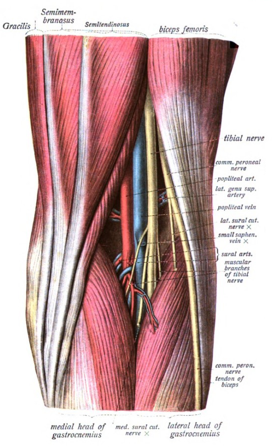


Popliteal Fossa Wikipedia


Types Of Popliteal Artery Entrapment Syndrome Paes I The Artery Download Scientific Diagram


1
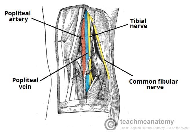


The Popliteal Fossa Borders Contents Teachmeanatomy


Popliteal Fossa Boundaries Contents And Applied Aspects Anatomy Qa
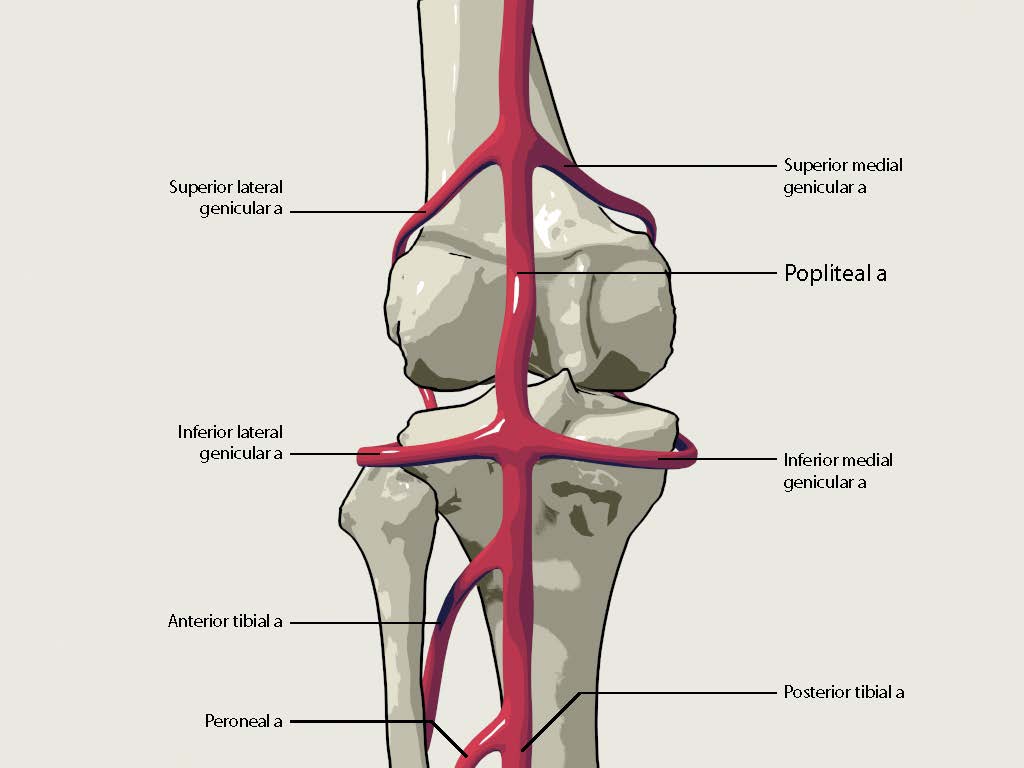


Popliteal Artery Anatomy And Course Bone And Spine


Popliteal Pulse What It Is And How To Find It


Venous Drainage Of Leg Anatomy Popliteal Artery And Vein Small Saphenous Vein Plantaris Muscle And Tendon In 21 Leg Anatomy Leg Vein Anatomy Human Body Muscles
/GettyImages-87313663-16bdfeaf37d048dbaef06b4f00b269b5.jpg)


Popliteal Vein Anatomy And Function


Anatomy Of The Popliteal Fossa Everything You Need To Know Dr Nabil Ebraheim Youtube
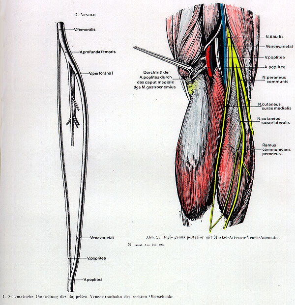


Anatomy Atlases Illustrated Encyclopedia Of Human Anatomic Variation Opus Ii Cardiovascular System Popliteal Artery And Vein Trapped In The Medial Head Of Gastrocnemius Variant Vein Duplicating The Popliteal Vein


Bats Better Anaesthesia Through Sonography
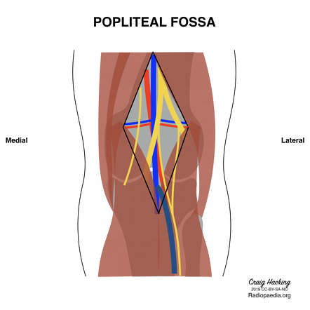


Popliteal Fossa Radiology Reference Article Radiopaedia Org


Anatomy Of The Knee Joint Doctor Stock
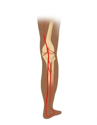


Popliteal Artery Entrapment Syndrome Frankel Cardiovascular Center Michigan Medicine


Http Www Kgmu Org Digital Lectures Medical Anatomy Lecture On Popliteal Fossa Pdf


Accessory Muscles Of The Knee Radsource


Popliteal Bypass Surgery Wikipedia


Popliteal Artery Anatomy And Course Bone And Spine
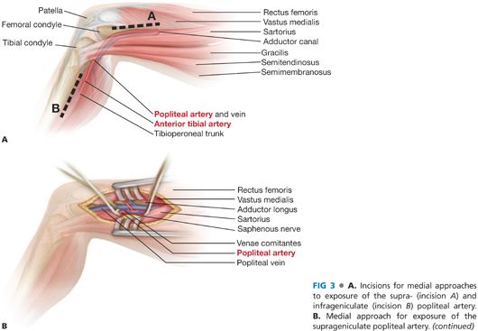


Surgical Exposure Of The Lower Extremity Arteries Thoracic Key


Popliteal Artery Entrapment Syndrome Wikipedia
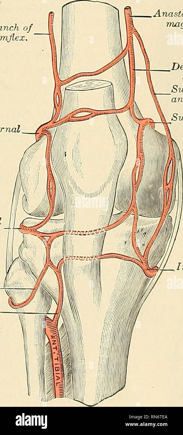


Popliteal Artery High Resolution Stock Photography And Images Alamy


Exercise Induced Leg Pain Due To Vascular Compression Syndrome Sems Journal


Endartery Stenosis Of The Popliteal Artery Mimicking Gastrocnemius Strain A Case Report1 Archives Of Physical Medicine And Rehabilitation


Http Www Kgmu Org Digital Lectures Medical Anatomy Lecture On Popliteal Fossa Pdf


Popliteal Artery Location Entrapment Popliteal Artery Aneurysm


Http Repository Tnmgrmu Ac In 1 bama Pdf
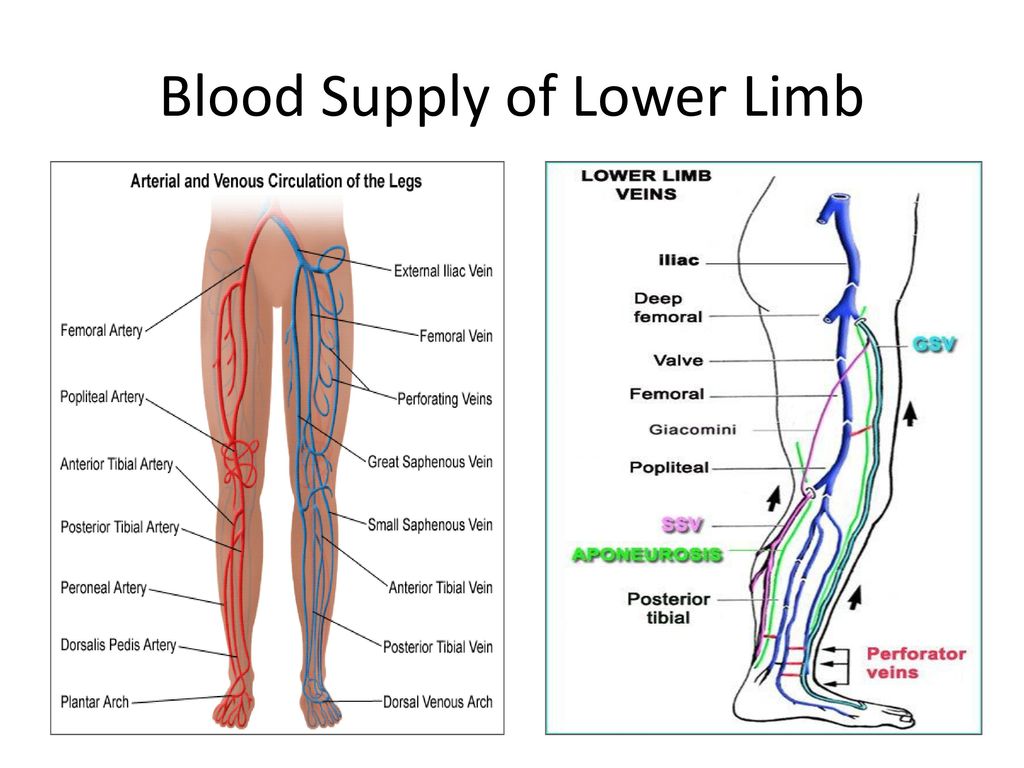


Blood Vessels Of Lower Limb Superior Inferior Gluteal Artery Femoral Popliteal Artery Science Online


Cambridge Orthopaedics Popliteal Block Uk


Anatomy Of The Popliteal Fossa Everything You Need To Know Dr Nabil Ebraheim Youtube
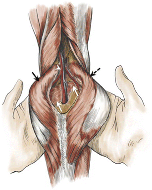


Uncommon Diseases Of The Popliteal Artery A Pictorial Review Insights Into Imaging Full Text
:background_color(FFFFFF):format(jpeg)/images/article/en/popliteal-artery/4fcNkYGQccOl0OkvY8cuw_gA4gQRyxVpboWSaGf9cUxw_A._poplitea_m01.png)


Popliteal Artery Anatomy Branches Location And Course Kenhub
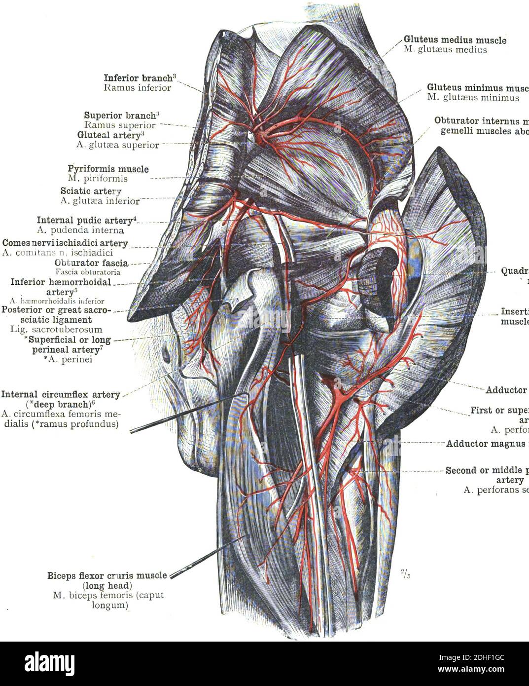


The Anatomy Of Popliteal Artery On A White Background Stock Photo Alamy


Popliteal Artery Anatomy And Course Bone And Spine


Which Structure Is Highlighted Popliteal Artery O Chegg Com
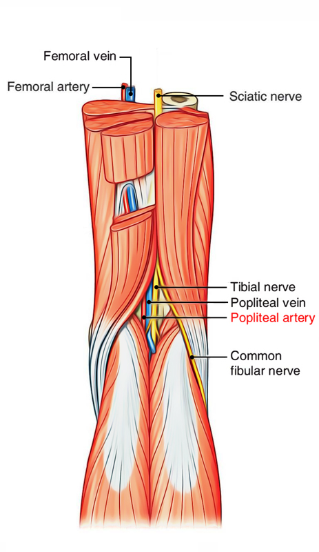


Easy Notes On Popliteal Artery Learn In Just 4 Minutes Earth S Lab


Popliteal Artery Artwork Stock Image C021 2101 Science Photo Library


01x Position Of Popliteal Vessels During Flexion Anatomy Exhibits


The Popliteal Artery Human Anatomy


Untitled Document


No comments:
Post a Comment